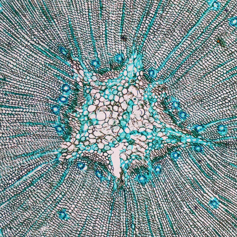
Wood Microscope . light and electron microscopy have contributed significantly to revolutionize our understanding of wood. under the microscope, any wood shows a number of distinctive characteristics determined by the growth pattern of the tree which produced it. Using light and electron microscopes to examine thin sections of wood, kew’s wood anatomists study the structure of individual cells and their arrangement within stems and roots. Cut samples to a radial thickness of 1cm. the microscope reveals that wood is composed of minute units called cells. wood must be observed under the optical microscope in very thin slices, called sections. The basic cell types are called tracheids, vessel members, fibres, and parenchyma. The sections can be easily obtained with a razor. Softwoods are made of tracheids and parenchyma, and.
from www.dreamstime.com
The basic cell types are called tracheids, vessel members, fibres, and parenchyma. Cut samples to a radial thickness of 1cm. the microscope reveals that wood is composed of minute units called cells. Softwoods are made of tracheids and parenchyma, and. light and electron microscopy have contributed significantly to revolutionize our understanding of wood. wood must be observed under the optical microscope in very thin slices, called sections. under the microscope, any wood shows a number of distinctive characteristics determined by the growth pattern of the tree which produced it. Using light and electron microscopes to examine thin sections of wood, kew’s wood anatomists study the structure of individual cells and their arrangement within stems and roots. The sections can be easily obtained with a razor.
Pine Wood micrograph stock image. Image of wood, macro 47393573
Wood Microscope under the microscope, any wood shows a number of distinctive characteristics determined by the growth pattern of the tree which produced it. Softwoods are made of tracheids and parenchyma, and. under the microscope, any wood shows a number of distinctive characteristics determined by the growth pattern of the tree which produced it. Cut samples to a radial thickness of 1cm. The sections can be easily obtained with a razor. the microscope reveals that wood is composed of minute units called cells. wood must be observed under the optical microscope in very thin slices, called sections. The basic cell types are called tracheids, vessel members, fibres, and parenchyma. Using light and electron microscopes to examine thin sections of wood, kew’s wood anatomists study the structure of individual cells and their arrangement within stems and roots. light and electron microscopy have contributed significantly to revolutionize our understanding of wood.
From www.scientificsonline.com
VictorianInspired Wood Microscope Kit Wood Microscope wood must be observed under the optical microscope in very thin slices, called sections. The basic cell types are called tracheids, vessel members, fibres, and parenchyma. the microscope reveals that wood is composed of minute units called cells. Using light and electron microscopes to examine thin sections of wood, kew’s wood anatomists study the structure of individual cells. Wood Microscope.
From www.dreamstime.com
Transverse Section of Pine Wood Under Microscope Stock Image Image of Wood Microscope under the microscope, any wood shows a number of distinctive characteristics determined by the growth pattern of the tree which produced it. The sections can be easily obtained with a razor. Softwoods are made of tracheids and parenchyma, and. the microscope reveals that wood is composed of minute units called cells. wood must be observed under the. Wood Microscope.
From microscope-microscope.org
Microscopy Wood Microbus Microscope Educational site Wood Microscope Using light and electron microscopes to examine thin sections of wood, kew’s wood anatomists study the structure of individual cells and their arrangement within stems and roots. light and electron microscopy have contributed significantly to revolutionize our understanding of wood. wood must be observed under the optical microscope in very thin slices, called sections. Softwoods are made of. Wood Microscope.
From www.dreamstime.com
Pine Wood Under the Microscope Stock Photo Image of microscope Wood Microscope The sections can be easily obtained with a razor. Using light and electron microscopes to examine thin sections of wood, kew’s wood anatomists study the structure of individual cells and their arrangement within stems and roots. under the microscope, any wood shows a number of distinctive characteristics determined by the growth pattern of the tree which produced it. Softwoods. Wood Microscope.
From tiffanycalum.blogspot.com
15+ Wood Under A Microscope TiffanyCalum Wood Microscope light and electron microscopy have contributed significantly to revolutionize our understanding of wood. Using light and electron microscopes to examine thin sections of wood, kew’s wood anatomists study the structure of individual cells and their arrangement within stems and roots. Softwoods are made of tracheids and parenchyma, and. wood must be observed under the optical microscope in very. Wood Microscope.
From microscope-microscope.org
Microscopy Wood Microbus Microscope Educational site Wood Microscope light and electron microscopy have contributed significantly to revolutionize our understanding of wood. the microscope reveals that wood is composed of minute units called cells. wood must be observed under the optical microscope in very thin slices, called sections. Softwoods are made of tracheids and parenchyma, and. under the microscope, any wood shows a number of. Wood Microscope.
From www.dreamstime.com
Pine Wood micrograph stock photo. Image of micro, optical 42350214 Wood Microscope Using light and electron microscopes to examine thin sections of wood, kew’s wood anatomists study the structure of individual cells and their arrangement within stems and roots. The sections can be easily obtained with a razor. under the microscope, any wood shows a number of distinctive characteristics determined by the growth pattern of the tree which produced it. Cut. Wood Microscope.
From www.dreamstime.com
Transverse Section Of Pine Wood Under Microscope Stock Photo Image of Wood Microscope Cut samples to a radial thickness of 1cm. Using light and electron microscopes to examine thin sections of wood, kew’s wood anatomists study the structure of individual cells and their arrangement within stems and roots. the microscope reveals that wood is composed of minute units called cells. under the microscope, any wood shows a number of distinctive characteristics. Wood Microscope.
From www.researchgate.net
4. Scanning electron microscope photograph of wood sections. Download Wood Microscope Softwoods are made of tracheids and parenchyma, and. Cut samples to a radial thickness of 1cm. The basic cell types are called tracheids, vessel members, fibres, and parenchyma. under the microscope, any wood shows a number of distinctive characteristics determined by the growth pattern of the tree which produced it. wood must be observed under the optical microscope. Wood Microscope.
From www.dreamstime.com
Ancient wooden microscope stock image. Image of college 107945127 Wood Microscope The sections can be easily obtained with a razor. Softwoods are made of tracheids and parenchyma, and. Cut samples to a radial thickness of 1cm. the microscope reveals that wood is composed of minute units called cells. under the microscope, any wood shows a number of distinctive characteristics determined by the growth pattern of the tree which produced. Wood Microscope.
From www.scientificsonline.com
VictorianInspired Wood Microscope Kit Wood Microscope Cut samples to a radial thickness of 1cm. wood must be observed under the optical microscope in very thin slices, called sections. Softwoods are made of tracheids and parenchyma, and. under the microscope, any wood shows a number of distinctive characteristics determined by the growth pattern of the tree which produced it. the microscope reveals that wood. Wood Microscope.
From arboretum.harvard.edu
Wood Under the Microscope Arnold Arboretum Arnold Arboretum Wood Microscope Using light and electron microscopes to examine thin sections of wood, kew’s wood anatomists study the structure of individual cells and their arrangement within stems and roots. the microscope reveals that wood is composed of minute units called cells. under the microscope, any wood shows a number of distinctive characteristics determined by the growth pattern of the tree. Wood Microscope.
From etsy.com
Bausch and Lomb Microscope with Wood Case patent 1908 Wood Microscope wood must be observed under the optical microscope in very thin slices, called sections. The sections can be easily obtained with a razor. the microscope reveals that wood is composed of minute units called cells. under the microscope, any wood shows a number of distinctive characteristics determined by the growth pattern of the tree which produced it.. Wood Microscope.
From www.pinterest.co.kr
Wood Sections Microscopic photography, Microscope art, Patterns in nature Wood Microscope The basic cell types are called tracheids, vessel members, fibres, and parenchyma. Cut samples to a radial thickness of 1cm. The sections can be easily obtained with a razor. Softwoods are made of tracheids and parenchyma, and. under the microscope, any wood shows a number of distinctive characteristics determined by the growth pattern of the tree which produced it.. Wood Microscope.
From microscope-microscope.org
Microscopy Wood Microbus Microscope Educational site Wood Microscope wood must be observed under the optical microscope in very thin slices, called sections. The sections can be easily obtained with a razor. the microscope reveals that wood is composed of minute units called cells. light and electron microscopy have contributed significantly to revolutionize our understanding of wood. Using light and electron microscopes to examine thin sections. Wood Microscope.
From fp.optics.arizona.edu
Wooden Culpeper Microscope Wood Microscope The basic cell types are called tracheids, vessel members, fibres, and parenchyma. the microscope reveals that wood is composed of minute units called cells. The sections can be easily obtained with a razor. Using light and electron microscopes to examine thin sections of wood, kew’s wood anatomists study the structure of individual cells and their arrangement within stems and. Wood Microscope.
From arboretum.harvard.edu
Wood Under the Microscope Arnold Arboretum Arnold Arboretum Wood Microscope Cut samples to a radial thickness of 1cm. The sections can be easily obtained with a razor. under the microscope, any wood shows a number of distinctive characteristics determined by the growth pattern of the tree which produced it. light and electron microscopy have contributed significantly to revolutionize our understanding of wood. The basic cell types are called. Wood Microscope.
From www.dreamstime.com
521 Wood Microscopic Photos Free & RoyaltyFree Stock Photos from Wood Microscope Softwoods are made of tracheids and parenchyma, and. light and electron microscopy have contributed significantly to revolutionize our understanding of wood. Cut samples to a radial thickness of 1cm. Using light and electron microscopes to examine thin sections of wood, kew’s wood anatomists study the structure of individual cells and their arrangement within stems and roots. The basic cell. Wood Microscope.
